Radiology is a medical specialization that utilizes imaging to diagnose and treat diseases in the body. In our hospital, a variety of diagnostic (therapeutic) and interventional radiology techniques are provided through state-of-the-art technological devices.
Diagnostic and therapeutic imaging techniques for the diagnosis and/or treatment of diseases include X-Ray (Radiography), ultrasonography, color Doppler ultrasonography, mammography, digital fluoroscopy, computed tomography (CT), and magnetic resonance imaging (MRI).
Some of the therapeutic radiology techniques used in our hospital include:
X-Ray (Radiography): X-ray produces an image of the internal structures of the body by exposing it to controlled X-rays. The images are typically recorded on a digital sensor and displayed on a computer screen.
Ultrasonography: Ultrasonography uses high-frequency sound waves emitted by an ultrasound probe to image the body. These waves travel through the body, reflecting off various tissue layers. The probe then detects these reflected waves, which are transferred to a screen for interpretation.
Computed Tomography (CT): CT is another X-ray technique that allows radiologists to visualize images in two-dimensional or three-dimensional forms. Whole-body CT scans can be performed within a few seconds and often require the use of contrast material to enhance image quality.
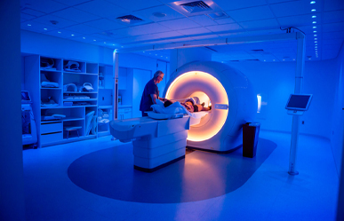
Magnetic Resonance Imaging (MRI): MRI uses magnetism and radio waves to create images similar to CT scans. It utilizes these techniques to generate a series of sectional images. MRI provides additional information about the characteristics of tissues and organs not easily discernible with other techniques. The acquisition of MRI images takes longer than traditional X-rays or CT scans.
Digital Fluoroscopy: Digital fluoroscopy involves using a series of low-dose X-rays to create a moving image of the body. It enables real-time visualization of organs and facilitates interventional procedures under imaging guidance.
Mammography: Mammography is a technique that uses X-rays to examine the breast for irregularities or early signs of cancer. Regular mammography screening for breast cancer is recommended for women starting from the age of 40.
Interventional radiology involves a set of techniques relying on radiological imaging guidance to precisely target treatment. These techniques include X-ray, fluoroscopy, ultrasonography, CT, and MRI.
Interventional radiology techniques often involve invasive procedures such as inserting a needle, cannula (tube), catheter, or wire into the patient for diagnosis and/or treatment. Procedures include angioplasty (placing a balloon into a blood vessel to widen and improve circulation), stenting (inserting a tube to keep a blood vessel open), and biopsies of organs like the lungs, breasts, kidneys, liver, and bones.
The well-known advantages of these minimally invasive techniques include reduced risks, shorter hospital stays, lower costs, increased comfort, and faster return to work.
In our hospital, all radiological diagnostic and treatment procedures for our patients are performed using the range of equipment listed above. All radiological examinations are made visible through the PACS system, can be stored for many years, and can be evaluated by doctors in various specialties through digital means on monitors.
The Radiology Department of Tekden Hospital is a unit equipped with a team of experts and state-of-the-art equipment, providing imaging and diagnostic services. Here are the detailed services offered in our department:
Imaging Techniques:
Computed Tomography (CT): Detailed cross-sectional images are obtained within the body using advanced technology devices.
Magnetic Resonance Imaging (MRI): Organs and tissues are visualized in detail using high magnetic fields.
Our MRI equipment is one of the best and most spacious MRI devices in Kayseri in terms of both quality and comfort.
X-rays: Static images of the skeleton and internal organs are taken.
Ultrasonography: Real-time imaging of organs using high-frequency sound waves.
Mammography and Imaging:
Imaging using digital mammography devices for breast cancer screening.
Additional evaluations are performed using breast ultrasound and magnetic resonance imaging.
Radiological Diagnosis and Reporting:
Evaluation of imaging results in detail by expert radiologists for diagnosis and reporting.
Coordination with relevant specialists for diagnosis and treatment.
Vascular and Cardiac Imaging:
Detailed examination of the vascular system and heart vessels.
Performance of special procedures such as angiography and coronary angiography.
Pediatric Radiology:
Imaging services specifically designed for pediatric radiological issues, conducted by specialists trained in pediatric radiology.
Dose control and the creation of a child-friendly imaging environment.
Tekden Hospital's Radiology Department provides the highest standards in diagnostic imaging with a patient-centered approach, cutting-edge technology, and an expert team. It works effectively with a focus on patient satisfaction and the early diagnosis and effective treatment of diseases.
