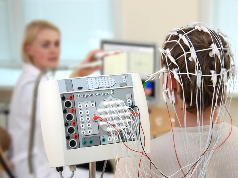
The technique that involves analyzing the electrical activity recorded from the scalp to demonstrate the distribution of different frequency waves on the brain and provide indirect information about the functioning of the brain is known as Quantitative Electroencephalography (QEEG). When repeated after treatment, QEEG can show the positive changes achieved through the treatment. As seen in the profiles obtained before and after treatment in the examples, QEEG allows observation of the correction of irregularities in brain chemistry through bioelectrical activity recording.
Understanding and monitoring brain function is crucial for effective treatment, especially in the case of many mental disorders being associated with brain diseases. In the treatment of depression, monitoring both the psychological or social aspects along with the biological dimension is particularly valuable and prioritized, especially in cases of treatment resistance. We are pleased to bring innovation in this regard at Tekden Hospital.
Measuring brain functions in adults, adolescents, and children to guide treatment is a desired and targeted goal in psychiatry. QEEG allows the measurement of bioelectrical activity, the end product of biological processes in the brain. Numerous validity and reliability studies have been conducted in this regard, making it one of the most usable methods among existing biological indicators.
Conventional and Quantitative EEG in Psychiatry New brain imaging techniques provide information about structural and functional abnormalities in various psychiatric disorders. These imaging techniques include magnetic resonance imaging, positron emission tomography of regional cerebral metabolic rate, cerebral regional blood flow, radioligand binding to neurotransmitter receptors, single-photon emission computed tomography (SPECT), functional magnetic resonance imaging (fMRI), magnetoencephalography, quantitative EEG (QEEG), event-related potentials (ERP), topographic QEEG, statistical probability mapping, and low-resolution electromagnetic tomography.
These brain imaging methods have shown that there is a clear association between mental illnesses and brain dysfunction. QEEG and ERP are non-invasive, sensitive tools with higher resolution compared to other methods, making them valuable in quantitatively evaluating the resting and stimulated activities of the brain in psychiatric practice.
Anxiety disorders, depression, dementia, obsessive-compulsive disorder, schizophrenia, learning difficulties, attention-deficit disorders with or without hyperactivity, and other disorders have been shown to involve brain dysfunction or an interaction between environmental factors and impaired neuroanatomic structures. Medications that alter the suitability of neurotransmitters and affect a hypothesized pathophysiology are routinely used in psychiatric practice. However, biological assessment methods are often not used for treatment selection and evaluation of physiological effects and effectiveness in cases of treatment-resistant conditions.
Why Should EEG be Used in Psychiatry Patients? To make a psychiatric diagnosis, physical or neurological conditions should first be ruled out. EEG abnormalities are considered in 64% - 68% of psychiatric patients, and this result carries more than eliminating organic brain damage, having diagnostic significance. Such EEG studies can assist in differential diagnosis, treatment selection, and evaluation. Some follow-up studies have shown that the initially examined QEEG profiles can distinguish groups among patients with the same DSM diagnosis, indicating different progressions of the disease or groups responding better to different areas.
EEG in Schizophrenia Various qualitative studies have reported EEG abnormalities in 20% to 60% of schizophrenic patients. A more specific finding for schizophrenia is a relatively low average alpha frequency, but some patients may show a rapid alpha rhythm. Catatonic patients often exhibit paroxysmal activity. When examining the EEG records of medicated and non-medicated schizophrenic patients, a decrease in alpha dominance and impaired alpha average frequency or decreased alpha response has been repeatedly reported. Increased beta activity has also been reported in many studies. Neuroleptics typically increase alpha dominance and decrease beta dominance. This suggests that some disorders in the EEG of medicated patients may be concealed.
In elderly schizophrenic patients, an increase in fast theta activity (7-7.5 Hz) is observed. Increased delta activity in the frontal areas is termed hypofrontality.
Various relationships with clinical conditions have also been reported:
However, several different EEG profiles can be observed in the schizophrenic patient population. When addressing this heterogeneity in research, four types of QEEG clusters have been identified. These clusters include delta and theta excess, theta excess, beta deficiency with theta and alpha excess, and alpha and beta excess. Previously untreated patients can exhibit three of these subtypes. Patients in different subtypes have been shown to respond differently to haloperidol and risperidone treatments.
Asymmetry findings in schizophrenia can vary in study results. While dominance in frontal areas is higher in the right hemisphere, it is higher in the posterior areas of the left hemisphere. Increased delta activity in the left anterior temporal area has been reported to differentiate schizophrenic patients from healthy controls.
Conclusion: The EEG and QEEG literature in schizophrenia becomes complicated due to the heterogeneous nature of the disease and the differences in drug histories and doses. However, despite these confusing factors, there are some recurring findings. The most frequently reported anomalies are an increase in delta and/or theta in frontal areas, a decrease in average frequency and power in the alpha band, and an increase in beta power. An increase in coherence in the frontal areas has also been frequently reported. Coherence measurements can contribute to distinguishing bipolar disorder from schizophrenia, as reduced coherence in the frontal areas has been reported to help differentiate schizophrenia from depression.
In summary, EEG and QEEG studies have provided valuable insights into the neurophysiological aspects of schizophrenia. The heterogeneity of EEG findings in schizophrenia suggests that different subtypes may exist within the disorder, and these subtypes may respond differently to pharmacological treatments. Incorporating QEEG into clinical practice may contribute to more personalized and effective treatment approaches for schizophrenia.